Baker's Cyst
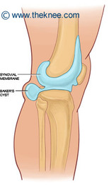
A Baker's Cyst is a swelling of the gastrocnemius-semimebranosus bursa in the medial popliteal fossa just distal to the crease in the knee. It often occurs as a complication of joint pathology, such as knee joint derangement or osteoarthritis. Baker's Cysts are often small and asymptomatic, but when they become symptomatic they are characterized by posterior knee pain, knee stiffness, and swelling or a mass behind the knee, especially when the knee is in extension. Symptoms may be worse with knee in hyperflexion, in standing and with activity.
If a Baker's Cyst ruptures, sometimes following strenuous exercise, the complications are painful and can mimic a DVT, with symptoms including warmth, tenderness and redness in the calf. Ecchymosis may be seen radiating distally to the medial malleolus, and proximally into the thigh. Rupture can cause posterior tibial nerve entrapment, anterior or posterior compartment syndrome, and occlusion of the popliteal artery with resulting lower leg ischemia. Baker's Cyst rupture is rare, but when it occurs the patient should promptly seek medical attention to decrease risk of permanent tissue damage.
Identifying Baker's Cyst
With the patient standing with knee in full extension, a mass maybe able to be palpated in the medial popliteal fossa. It may soften or disappear as the knee moves to 45 degrees flexion. Radiography and ultrasound can be used for further diagnosis, although ultrasound is more effective in visualizing this fluid-filled area.
How symptoms differ from DVT:
Presence of knee pain and joint swelling and inflammatory joint disease are not commonly associated with DVT but will most likely be found with a Baker's Cyst.
Helfgott, S. (2013) "Popliteal (Baker's) Cyst" UpToDate.
