Peripheral Vascular Disease
Peripheral Arterial Disease
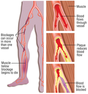
Peripheral Arterial Disease (PAD) is a manifestation of systemic atherosclerosis that is often asymptomatic. The patient with PAD is at increased risk for coronary artery disease and cerebrovascular disease, unfortunately these patients are often under-identified and under-treated. The skin may appear shiny, thin and pale, with thick nails on the affected extremity and hair loss. There may be ulcers on the ankle or foot associated with arterial insufficiency.
How PAD may mimic DVT
http://www.racgp.org.au/afp/2013/june/peripheral-arterial-disease-diagnosis/
Peripheral Arterial Disease (PAD) is a manifestation of systemic atherosclerosis that is often asymptomatic. The patient with PAD is at increased risk for coronary artery disease and cerebrovascular disease, unfortunately these patients are often under-identified and under-treated. The skin may appear shiny, thin and pale, with thick nails on the affected extremity and hair loss. There may be ulcers on the ankle or foot associated with arterial insufficiency.
How PAD may mimic DVT
- Patient may experience intermittent claudication: Calf pain with exertion
- Ankle-Brachial Index
- Look for rubor in feet in dependent position vs elevated position
http://www.racgp.org.au/afp/2013/june/peripheral-arterial-disease-diagnosis/
Venous Insufficiency
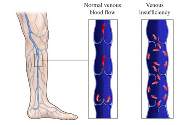
Venous Insufficiency involves failure of the venous valves. Arterial pulse pressure will be normal, and skin color will be pink to cyanotic, with potential brown pigmenting at the ankle. The skin may be discolored or scaly from stasis dermatitis, ulcers on the ankles, toes or fingers, and varicose veins. The patient may experience increased pain in standing as more blood pools in the lower extremities, and the pain may be relieved with lower extremity elevation or support hose.
Screening tools: Capillary Refill Time increased, ABI will be normal
