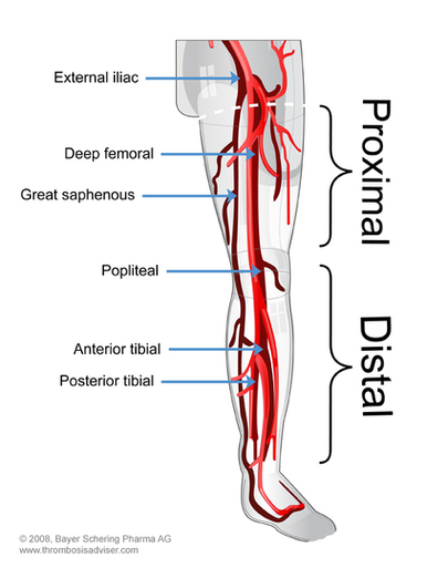
Lower Extremity Vascular Anatomy
Lower Extremity DVT's
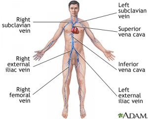
The most common sites for DVT's are in the popliteal, femoral, and iliac veins. They can also be found, less commonly, in the posterior tibial and short saphenous vein. When an embolus breaks free, it can travel from either the posterior tibial or short saphenous vein, into the popliteal vein, then to the femoral vein, external iliac, common iliac, and then into the inferior vena cava. From here, the embolus can enter the heart and lungs.
The most common sites for DVT's are in the popliteal, femoral, and iliac veins. They can also be found, less commonly, in the posterior tibial and short saphenous vein. When an embolus breaks free, it can travel from either the posterior tibial or short saphenous vein, into the popliteal vein, then to the femoral vein, external iliac, common iliac, and then into the inferior vena cava. From here, the embolus can enter the heart and lungs.
References
- Gilroy AM, MacPherson BR, Ross LM. Atlas of Anatomy. Thieme; 2008, (422-423).
- Riddle DL, Wells PS. Diagnosis of lower extremity deep vein thrombosis in outpatients. Physical Therapy. 2004 Aug;84(8):729-35.
